Image fraud in life sciences is a critical challenge. Manipulated images in research questions the credibility of research, waste valuable resources, and, in some cases, lead to harmful outcomes in medical and biological advancements. This article explores the scope of image fraud, its impact, the detection technologies available, and strategies to mitigate it.
Understanding Image Fraud in Life Sciences
Image fraud involves the deliberate alteration or manipulation of visual data to misrepresent findings. A study found that out of 20,621 papers published in 40 scientific journals from 1995 to 2014, 3.8% of images were problematic, with almost half of these cases being unintentional. Image fraud, whether intentional and unintentional can erode scientific integrity and trust, lead to retractions, and damage the credibility of journals, institutions, and researchers involved. Furthermore, it wastes funding and time and may produce to incorrect results leading to ineffective or harmful medical treatments.
Here are some types of image fraud and their impacts:
1. Image Duplication:
What?
The same image is reused within a paper or across different studies to support multiple claims.
Impact
Creates a false impression of consistency, leading to misguided conclusions and potentially influencing follow-up research or clinical trials.
Example
A study in oncology was retracted after it was discovered that identical microscopy images were used to represent different experimental conditions.
2. Image Splicing:
What?
Fragments of the same or different images are pasted together to fabricate a single cohesive image.
Impact
Misrepresents data, questioning data reliability, reproducibility, and credibility.
Example
Spliced Western blot bands can falsely indicate protein interactions, misguiding therapeutic research.
3. Image Enhancement:
What?
Adjustments such as altering brightness, contrast, color saturation, or sharpness beyond acceptable limits to emphasize or obscure features.
Impact
While subtle, such enhancements can distort data interpretation or pixelate images, especially in diagnostic or quantitative studies, leading to misinterpretation.
Example
Manipulated imaging in cell biology studies has led to exaggerated claims about cell proliferation rates, skewing experimental outcomes.
4. Image Cropping and Masking:
What?
Selective cropping to remove context or details that might refute the presented hypothesis.
Impact
This prevents a holistic understanding of the data, leading to biased interpretations and decisions.
Example
Cropped images hiding significant control data, which, when revealed, contradicts the study conclusions.
5. Mislabeling and Misrepresentation:
What?
Images are intentionally mislabeled or presented out of context.
Impact
Mislabeling misleads readers, causing cascading errors in downstream research.
Example
An immunohistochemistry image labeled as “control,” was later found to be a treated sample, misleading the study’s interpretation.
Given the diverse and pervasive nature of image fraud in life sciences, it is essential to equip the research community with robust tools and methodologies for detection. Advanced technologies and human oversight are pivotal in identifying these manipulations before they infiltrate the scientific record.
Tools and Techniques for Detecting Image Fraud
Advanced technologies and methods have been developed to detect image fraud effectively:
1. Forensic software:
Tools such as ImageJ, FotoForensics, and specialized software developed for academic purposes can detect inconsistencies. These tools analyze pixelation, contrast, and metadata to uncover evidence of duplication, splicing, or enhancement.
2. AI-driven analysis:
Artificial intelligence can help in identifying image duplications and manipulations. Machine learning models trained on large datasets can detect subtle anomalies, such as identical patterns in ostensibly different images. Furthermore, it can also detect duplication by scanning the images across the published resources.
3. Automated screening tools:
Journals and publishers increasingly use tools like Proofig and Similarity Check to automatically screen submitted images for signs of manipulation.
4. Human expertise:
Despite technological advancements, expert reviewers are often essential for identifying nuanced manipulations. Journals often assemble dedicated image integrity teams to review submissions manually.
Preventative Strategies
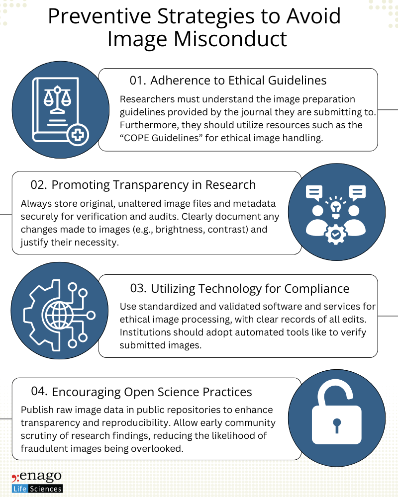
Proactive measures are essential to minimize image fraud and ensure research integrity. Institutions should prioritize education on ethical guidelines and acceptable image processing techniques. Researchers must understand the line between permissible adjustments (e.g., cropping, brightness adjustments) and unethical alterations. Additionally, publishers and journals must adopt stringent submission guidelines, including mandatory raw data submission and routine image screening. Also, they must encourage researchers to publish raw image data alongside processed results to promote transparency.

Image fraud in life sciences jeopardizes the foundation of scientific inquiry. Addressing this issue requires a combination of technological solutions, stringent policies, and cultural shifts toward ethical research practices. By leveraging advanced detection tools, enforcing transparent policies, and fostering a commitment to integrity, the scientific community can safeguard the reliability of its discoveries and reinforce public trust.
Author:

Anagha Nair
Editorial Assistant, Enago Academy
Medical Writer, Enago Life Sciences
Connect with Anagha on LinkedIn

